A Short Literature Review (on DR)
Diabetic Retinopathy (DR)
Diabetes mellitus is a condition in which the pancreas, a gland in the abdomen, produces insufficient quantity of a hormone called insulin. Due to a deficiency of insulin, the body is unable to control the level of sugar in blood. Diabetes can affect the eye sight in several ways. Diabetic patients have a higher likelihood of developing cataract, glaucoma and damage to the retina.
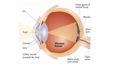
Retina is a light sensitive layer at the back of eye on which the images are created and from here transmitted to the brain. This layer has a fine mesh-work of very small blood vessels that get damaged in diabetes. This is characterized by a condition called diabetic retinopathy. All diabetic patients are at risk of developing diabetic retinopathy. The longer the patient has diabetes and the more uncontrolled it is, the greater the risk.
In diabetic retinopathy the small blood vessels of retina become weakened and develop small outpouchings called microaneurysms. These vessels also become more permeable that its proteins and other substances readily leak out through walls of the blood vessels. In the later stages, these blood vessels may get completely blocked. Due to the diseased vessels, the retina is deprived of adequate oxygen supply and this stimulates growth of new, abnormal vessels in the retina.
SYMPTOMS
Patients may remain free of symptoms in the
early phases of the disease. A problem is first noted when the abnormal vessels
bleed into the jelly of the eye called vitreous. This phenomenon is called
vitreous haemorrhage. This presents like noticing black spots in the vision that
are floating or as cobwebs. There may a loss of vision that might be sudden or
gradual. The bleeding into vitreous leads to development of critical scar that
pulls on to the retina causing it to detach from the eyeball (Retinal Detachment).
DIAGNOSIS
An eye specialist needs to do a thorough
examination. All diabetic patients must visit an ophthalmologist once every
year for an eye check-up to detect any abnormality in its early, treatable
phase. The doctor may also perform a test called Fluorescein Angiography in which a dye is injected into a vein in the arm and photographs
of the eye are taken using a special camera. This test helps to detect an abnormal
leakage of fluid in the eye, blockage in the blood vessels of the retina and
presence of new abnormal blood vessels in the retina.
A NORMAL EYE
TREATMENT
Several treatment modalities are available depending on the extent of damage. Laser Photocoagulation is a technique in which the abnormal leakage of fluid can be reduced using Laser. This helps to regress the abnormal blood vessels in the retina, thereby reducing the chances of bleeding. This treatment does not improve one’s vision, but just helps to maintain it at that level and slows the progression of the fall in vision. In some cases, the blood in the vitreous and the abnormal scar tissue may need to be removed by a procedure called Vitrectomy. Some techniques like vitreous microsurgery and endolaser have shown an improvement in majority of cases.